Neurological Conditions
Neurology refers to the diagnosis and treatment of disorders and diseases of the nervous system, which includes the spinal cord, brain and nerves. Neurologists can often diagnose and treat muscle disorders as well. Neurological disorders are somewhat common and can range from severe life-threatening conditions like stroke, Alzheimer’s and meningitis to less harmful but almost always debilitating conditions, such as migraine, epilepsy and sleep disorders.
Headache/Migraine
Migraines
Specific treatment for headaches will be determined by us based on:
- Your age, overall health, and medical history
- Severity and frequency of the symptoms
- Your tolerance for specific medications, procedures, or therapies
- Your opinion or preference
Treatment may include:
- Drug therapy (medication)
- Stress reduction
- Dietary evaluation
- Regular exercise
- Cold packs applied to the skin
- Pressure applied to the head
- Biofeedback training
The ultimate goal of treatment is to stop headaches from occurring. Adequate headache management depends on the accurate identification of the type of headache and may include:
- Avoiding known triggers, such as certain foods and beverages, lack of sleep, and fasting
- Changing eating habits
- Exercise
- Resting in a quiet, dark environment
- Medications, as recommended by your doctor
- Stress management
Migraine headaches may require specific medication management including:
- Abortive medications. Medications, prescribed by your doctor, that act on specific receptors in blood vessels in the head and can stop a headache in progress.
- Rescue medications. Medications purchased over-the-counter, such as analgesics (pain relievers), to stop the headache.
- Preventive medications. Medications, prescribed by your doctor, that are taken daily to reduce the onset of severe migraine headaches.
Successful prevention has come from many drug classes, including beta blockers, antidepressants, and anti-seizure drugs, as well as pain relievers. However, Botox is the first FDA-approved preventive therapy for chronic migraine headaches. Regular injections to the forehead and neck with Botox prevents migraines before they even start.
Some migraines may require immediate medical attention, including hospitalization for observation, diagnostic testing, or even surgery. Treatment is individualized, depending on the severity and frequency of symptoms.
“To make an appointment, call 727-712-1567. If you are experiencing a life threatening condition, please call 911.”
Lower Back Pain
Lower Back Pain
Every back pain and neck pain patient is unique, with different degrees of problems associated with a bone or disc abnormality. A neurologist is trained to discover the causes of symptoms, as well as using EMG testing to assess the injury to nerves and whether it is reversible in the short and long term.
Specific treatment for low back pain will be determined by your doctor based on:
- Your age, overall health, and medical history
- Diagnosis
- Extent of the condition
- Your tolerance for specific medications, procedures, or therapies
- Expectations for the course of the condition
- Your opinion or preference
Treatment may include:
- Activity modification
- Medication
- Physical rehabilitation and/or therapy
- Occupational therapy
- Weight loss (if overweight)
- No smoking
- Following a prevention program (as directed by your doctor)
- Surgery
- Assistive devices (for example, mechanical back supports)
Low back pain rehabilitation
Generally, there are 3 phases to low back pain rehabilitation. These include the following:
- Phase I: Acute Phase. During this initial phase, the physiatrist and treatment team focus on making a diagnosis, developing an appropriate treatment plan, and implementing the treatment regimen to reduce the initial low back pain and source of inflammation. This may include any or all of the items listed above and/or the utilization of ultrasound, electrical stimulation, or specialized injections.
- Phase II: Recovery Phase. Once the initial pain and inflammation are better managed, the rehabilitation team then focuses on helping the patient to restore working function of the body. This includes returning the patient to normal daily activities while implementing a specialized exercise program that is designed to help the individual regain flexibility and strength.
- Phase III: Maintenance Phase. The goal of this phase of low back pain rehabilitation is 2-fold — educating the individual on ways to prevent further injury and strain to the back, and helping the individual to maintain an appropriate level of physical fitness to help further increase strength and endurance.
“To make an appointment, call 727-712-1567. If you are experiencing a life threatening condition, please call 911.”
Numbness / Tingling
Numbness/Tingling and Peripheral Neuropathy
Peripheral Neuropathy
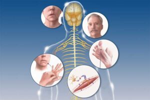
Peripheral neuropathy is a type of damage to the nervous system. Specifically, it occurs when there is a problem with your peripheral nervous system, the network of nerves that transmits information from your central nervous system (your brain and spinal cord) to the rest of your body.
The symptoms of peripheral neuropathy can vary greatly depending on what part of the body is affected. Symptoms can range from tingling or numbness in a certain body part to more serious effects such as burning pain or paralysis.
Treatment
Usually a peripheral neuropathy can’t be cured, but you can do a lot of things to prevent it from getting worse. If an underlying condition like diabetes is at fault, your doctor will treat that first and then treat the pain and other symptoms of neuropathy.
In some cases, over-the-counter (OTC) pain relievers can help. Other times, prescription drugs are needed. Some of these drugs include mexiletine, a medication developed to correct irregular heart rhythms; antiepileptic drugs, such as gabapentin, phenytoin, and carbamazepine; and some classes of antidepressants, including tricyclics such as amitriptyline.
Lidocaine injections and patches may help with pain in other instances. And in extreme situations, surgery can be used to destroy nerves or repair injuries that are causing neuropathic pain and symptoms.
“To make an appointment, call 727-712-1567. If you are experiencing a life threatening condition, please call 911.”
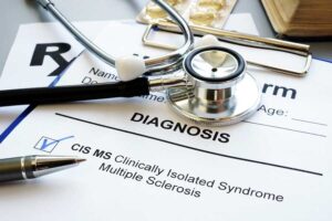
Multiple Sclerosis (MS)
Multiple Sclerosis (MS)
Specific treatment for MS will be determined by your doctor based on:
- Your age, overall health, and medical history
- Extent of the disease
- Your tolerance for specific medications, procedures, or therapies
- Expectations for the course of the disease
- Your opinion or preference
Treatments for the conditions associated with MS may include the following:
- Medication
- Clinical trials
- Assistive technology
- Rehabilitation activities
There is no cure yet for MS. However, there are strategies to modify the disease course, treat exacerbations, manage symptoms, and improve function and mobility.
Rehabilitation for people with MS
Rehabilitation varies depending on the range, expression, severity, and progression of symptoms. MS rehabilitation may help to accomplish the following:
- Restore functions that are essential to the activities of daily living (ADLs)
- Help the patient to reach maximum independence
- Promote family involvement
- Empower the patient to make the appropriate decisions relating to his or her care
- Educate the patient regarding the use of assistive devices (for example, canes, braces, or walkers)
- Establish an appropriate exercise program that promotes muscle strength, endurance, and control
- Reestablish motor skills
- Improve communication skills for patients who have difficulty speaking because of weakness or incoordination of face and tongue muscles
- Manage bowel or bladder incontinence
- Provide cognitive retraining
- Adapt the home environment to emphasize function, safety, accessibility, and mobility
“To make an appointment, call 727-712-1567. If you are experiencing a life threatening condition, please call 911.”
Neck Pain
Neck Pain
Diagnosis
Your doctor will take a medical history and do an exam. He or she will check for tenderness, numbness and muscle weakness, as well as see how far you can move your head forward, backward and side to side.
Imaging tests
Your doctor might order imaging tests to get a better picture of the cause of your neck pain. Examples include:
- X-rays. X-rays can reveal areas in your neck where your nerves or spinal cord might be pinched by bone spurs or other degenerative changes.
- CT scan. CT scans combine X-ray images taken from many different directions to produce detailed cross-sectional views of the internal structures of your neck.
- MRI. MRI uses radio waves and a strong magnetic field to create detailed images of bones and soft tissues, including the spinal cord and the nerves coming from the spinal cord.
It’s possible to have X-ray or MRI evidence of structural problems in your neck without having symptoms. Imaging studies are best used as an adjunct to a careful history and physical exam to determine the cause of your pain.
Other tests
- Electromyography (EMG). If your doctor suspects your neck pain might be related to a pinched nerve, he or she might suggest an EMG. It involves inserting fine needles through your skin into a muscle and performing tests to measure the speed of nerve conduction to determine whether specific nerves are functioning properly.
- Blood tests. Blood tests can sometimes provide evidence of inflammatory or infectious conditions that might be causing or contributing to your neck pain.
“To make an appointment, call 727-712-1567. If you are experiencing a life threatening condition, please call 911.”
Alzheimer’s Disease / Dementia
Alzheimer’s Disease / Dementia
Specific treatment for Alzheimer’s disease will be determined by your doctor based on:
- Your age, overall health, and medical history
- Extent of the disease
- Your tolerance for specific medications, procedures, or therapies
- Expectations for the course of the disease
- Your opinion or preference
At this time, there is no cure for Alzheimer’s, no way of slowing down the progression of this disease, and no treatment available to reverse the deterioration of Alzheimer’s disease. New research findings give reason for hope, and several drugs are being studied in clinical trials to determine if they can slow the progress of the disease or improve memory for a period of time.
There are some medications available to assist in managing some of the most troubling symptoms of Alzheimer’s disease, including the following:
- Depression
- Behavioral disturbance
- Sleeplessness
In managing the disease, physical exercise and social activity are important, as are proper nutrition, health maintenance, and a calm and well-structured environment.
What are Alzheimer’s rehabilitation programs?
The rehabilitation program for people with Alzheimer’s differs depending on the symptoms, expression, and progression of the disease, and the fact that making a diagnosis of Alzheimer’s is so difficult. These variables determine the amount and type of assistance needed for the Alzheimer’s individual and family.
With Alzheimer’s rehabilitation, it is important to remember that, although any skills lost will not be regained, the caregiving team must keep in mind the following considerations:
- To manage the disease, plan a balanced program of physical exercise, social activity, proper nutrition, and health maintenance activities.
- Plan daily activities that help to provide structure, meaning, and accomplishment for the individual.
- As functions are lost, adapt activities and routines to allow the individual to participate as much as possible.
- Keep activities familiar and satisfying.
- Allow the individual to complete as many things by himself or herself as possible. The caregiver may need to initiate an activity, but allow the individual to complete it as much as he or she can.
- Provide “cues” for desired behavior (for example, label drawers, cabinets, and closets according to their contents).
- Keep the individual out of harm’s way by removing all safety risks (for example, car keys and matches).
- As a caregiver (full-time or part-time), understand your own physical and emotional limitations.
“To make an appointment, call 727-712-1567. If you are experiencing a life threatening condition, please call 911.”
Motor Neuron Disease / ALS
Motor Neuron Disease / ALS
What is Motor Neuron Disease?
Motor neuron disease is a progressive condition which occurs when certain nerve cells degenerate and die. There are two types of motor neuron cells. The upper motor neuron begins in the brain and ends in the spinal cord. The lower motor neuron starts in the spinal cord and ends in the muscles. Nerve cell degeneration and death causes muscle weakness, abnormal reflexes and decreased ability of the brain to control muscle movement. The affected nerve cells do not grow back, but healthy nerve cells can attempt to reconnect to the muscles, which slows the progression of the disease. There are several types of motor neuron disease, including amyotrophic lateral sclerosis (ALS or Lou Gehrig’s Disease), spinal muscular atrophy, Werdnig-Hoffman disease, and infantile spinal muscular atrophy. It only affects the skeletal muscles and does not affect automatic muscles like the heart.
Who gets Motor Neuron Disease?
Motor neuron disease can affect anyone, but most people are over the age of 40, and men are affected slightly more often than women. Approximately 2 in 100,000 people will get motor neuron disease. It does not seem to be caused by specific foods, lifestyles, or injuries. It is hereditary in some people (5-10%).
How is Motor Neuron Disease diagnosed?
After learning the patient’s history and performing a physical examination, a physician may perform an EMG study. They may also use an MRI or blood studies to look for other diseases which can present like a motor neuron disease.
How is Motor Neuron Disease treated?
There is no cure for motor neuron disease. Only one medication is currently available, called Riluzole. It slows the progression of ALS by a small amount. Physical therapy and bracing can also help with symptoms.
“To make an appointment, call 727-712-1567. If you are experiencing a life threatening condition, please call 911.”
Tremor / Shaking
Tremors / Shaking
Some people with essential tremor don’t require treatment if their symptoms are mild. But if your essential tremor is making it difficult to work or perform daily activities, discuss treatment options with your doctor.
Medications
- Beta blockers. Normally used to treat high blood pressure, beta blockers such as propranolol (Inderal) help relieve tremors in some people. Beta blockers may not be an option if you have asthma or certain heart problems. Side effects may include fatigue, lightheadedness or heart problems.
- Anti-seizure medications. Epilepsy drugs, such as primidone (Mysoline), may be effective in people who don’t respond to beta blockers. Other medications that might be prescribed include gabapentin (Gralise, Neurontin) and topiramate (Topamax, Qudexy XR). Side effects include drowsiness and nausea, which usually disappear within a short time.
- Tranquilizers. Doctors may use benzodiazepine drugs such as clonazepam (Klonopin) to treat people for whom tension or anxiety worsens tremors. Side effects can include fatigue or mild sedation. These medications should be used with caution because they can be habit-forming.
- OnabotulinumtoxinA (Botox) injections. Botox injections might be useful in treating some types of tremors, especially head and voice tremors. Botox injections can improve tremors for up to three months at a time.However, if Botox is used to treat hand tremors, it can cause weakness in your fingers. If Botox is used to treat voice tremors, it can cause a hoarse voice and difficulty swallowing.
Therapy
Doctors might suggest physical or occupational therapy. Physical therapists can teach you exercises to improve your muscle strength, control and coordination.
Occupational therapists can help you adapt to living with essential tremor. Therapists might suggest adaptive devices to reduce the effect of tremors on your daily activities, including:
- Heavier glasses and utensils
- Wrist weights
- Wider, heavier writing tools, such as wide-grip pens
Surgery
Surgery might be an option if your tremors are severely disabling and you don’t respond to medications.
- Deep brain stimulation. This is the most common type of surgery for essential tremor. It’s generally the preferred procedure in medical centers with significant experience in performing this surgery. Doctors insert a long, thin electrical probe into the portion of your brain that causes your tremors (thalamus). A wire from the probe runs under your skin to a pacemaker-like device (neurostimulator) implanted in your chest. This device transmits painless electrical pulses to interrupt signals from your thalamus that may be causing your tremors.Side effects of deep brain stimulation can include equipment malfunction; problems with motor control, speech or balance; headaches; and weakness. Side effects often go away after some time or adjustment of the device.
- Focused ultrasound thalamotomy. This noninvasive surgery involves using focused sound waves that travel through the skin and skull. The waves generate heat to destroy brain tissue in a specific area of the thalamus to stop a tremor. A surgeon uses magnetic resonance imaging to target the correct area of the brain and to be sure the sound waves are generating the exact amount of heat needed for the procedure.Focused ultrasound thalamotomy creates a lesion that can result in permanent changes to brain function. Some people have experienced altered sensation, trouble with walking or difficulty with movement. However, most complications go away on their own or are mild enough that they don’t interfere with quality of life.
“To make an appointment, call 727-712-1567. If you are experiencing a life threatening condition, please call 911.”
Parkinson’s
Parkinson's Disease
No specific test exists to diagnose Parkinson’s disease. Your doctor trained in nervous system conditions (neurologist) will diagnose Parkinson’s disease based on your medical history, a review of your signs and symptoms, and a neurological and physical examination.
Treatment
Parkinson’s disease can’t be cured, but medications can help control your symptoms, often dramatically. In some more advanced cases, surgery may be advised.
Your doctor may also recommend lifestyle changes, especially ongoing aerobic exercise. In some cases, physical therapy that focuses on balance and stretching also is important. A speech-language pathologist may help improve your speech problems.
Medications
Medications may help you manage problems with walking, movement and tremor. These medications increase or substitute for dopamine.
People with Parkinson’s disease have low brain dopamine concentrations. However, dopamine can’t be given directly, as it can’t enter your brain.
You may have significant improvement of your symptoms after beginning Parkinson’s disease treatment. Over time, however, the benefits of drugs frequently diminish or become less consistent. You can usually still control your symptoms fairly well.
Medications your doctor may prescribe include:
- Carbidopa-levodopa. Levodopa, the most effective Parkinson’s disease medication, is a natural chemical that passes into your brain and is converted to dopamine.Levodopa is combined with carbidopa (Lodosyn), which protects levodopa from early conversion to dopamine outside your brain. This prevents or lessens side effects such as nausea.Side effects may include nausea or lightheadedness (orthostatic hypotension).After years, as your disease progresses, the benefit from levodopa may become less stable, with a tendency to wax and wane (“wearing off”).Also, you may experience involuntary movements (dyskinesia) after taking higher doses of levodopa. Your doctor may lessen your dose or adjust the times of your doses to control these effects.
- Inhaled carbidopa-levodopa. Inbrija is a new brand-name drug delivering carbidopa-levodopa in an inhaled form. It may be helpful in managing symptoms that arise when oral medications suddenly stop working during the day.
- Carbidopa-levodopa infusion. Duopa is a brand-name medication made up of carbidopa and levodopa. However, it’s administered through a feeding tube that delivers the medication in a gel form directly to the small intestine.Duopa is for patients with more-advanced Parkinson’s who still respond to carbidopa-levodopa, but who have a lot of fluctuations in their response. Because Duopa is continually infused, blood levels of the two drugs remain constant.Placement of the tube requires a small surgical procedure. Risks associated with having the tube include the tube falling out or infections at the infusion site.
- Dopamine agonists. Unlike levodopa, dopamine agonists don’t change into dopamine. Instead, they mimic dopamine effects in your brain.They aren’t as effective as levodopa in treating your symptoms. However, they last longer and may be used with levodopa to smooth the sometimes off-and-on effect of levodopa.Dopamine agonists include pramipexole (Mirapex), ropinirole (Requip) and rotigotine (Neupro, given as a patch). Apomorphine (Apokyn) is a short-acting injectable dopamine agonist used for quick relief.Some of the side effects of dopamine agonists are similar to the side effects of carbidopa-levodopa. But they can also include hallucinations, sleepiness and compulsive behaviors such as hypersexuality, gambling and eating. If you’re taking these medications and you behave in a way that’s out of character for you, talk to your doctor.
- MAO B inhibitors. These medications include selegiline (Zelapar), rasagiline (Azilect) and safinamide (Xadago). They help prevent the breakdown of brain dopamine by inhibiting the brain enzyme monoamine oxidase B (MAO B). This enzyme metabolizes brain dopamine. Selegiline given with levodopa may help prevent wearing-off.Side effects of MAO B inhibitors may include headaches, nausea or insomnia. When added to carbidopa-levodopa, these medications increase the risk of hallucinations.These medications are not often used in combination with most antidepressants or certain narcotics due to potentially serious but rare reactions. Check with your doctor before taking any additional medications with an MAO B inhibitor.
- Catechol O-methyltransferase (COMT) inhibitors. Entacapone (Comtan) is the primary medication from this class. This medication mildly prolongs the effect of levodopa therapy by blocking an enzyme that breaks down dopamine.Side effects, including an increased risk of involuntary movements (dyskinesia), mainly result from an enhanced levodopa effect. Other side effects include diarrhea, nausea or vomiting.Tolcapone (Tasmar) is another COMT inhibitor that is rarely prescribed due to a risk of serious liver damage and liver failure.
- Anticholinergics. These medications were used for many years to help control the tremor associated with Parkinson’s disease. Several anticholinergic medications are available, including benztropine (Cogentin) or trihexyphenidyl.However, their modest benefits are often offset by side effects such as impaired memory, confusion, hallucinations, constipation, dry mouth and impaired urination.
- Amantadine. Doctors may prescribe amantadine alone to provide short-term relief of symptoms of mild, early-stage Parkinson’s disease. It may also be given with carbidopa-levodopa therapy during the later stages of Parkinson’s disease to control involuntary movements (dyskinesia) induced by carbidopa-levodopa.Side effects may include a purple mottling of the skin, ankle swelling or hallucinations.
“To make an appointment, call 727-712-1567. If you are experiencing a life threatening condition, please call 911.”
Aneurysm
Aneurysm
A brain aneurysm (AN-yoo-riz-um) is a bulge or ballooning in a blood vessel in the brain. It often looks like a berry hanging on a stem.
A brain aneurysm can leak or rupture, causing bleeding into the brain (hemorrhagic stroke). Most often a ruptured brain aneurysm occurs in the space between the brain and the thin tissues covering the brain. This type of hemorrhagic stroke is called a subarachnoid hemorrhage.
A ruptured aneurysm quickly becomes life-threatening and requires prompt medical treatment.
Most brain aneurysms, however, don’t rupture, create health problems or cause symptoms. Such aneurysms are often detected during tests for other conditions.
Treatment for an unruptured brain aneurysm may be appropriate in some cases and may prevent a rupture in the future. Talk with your caregiver to ensure you understand the best options for your specific needs.
“To make an appointment, call 727-712-1567. If you are experiencing a life threatening condition, please call 911.”
Stroke / TIA
Stroke/TIA
Once we have determined the cause of your transient ischemic attack, the goal of treatment is to correct the abnormality and prevent a stroke. Depending on the cause of your TIA, your doctor may prescribe medication to reduce the tendency for blood to clot or may recommend surgery or a balloon procedure (angioplasty).
Medications
Doctors use several medications to decrease the likelihood of a stroke after a transient ischemic attack. The medication selected depends on the location, cause, severity and type of TIA. Your doctor may prescribe:
- Anti-platelet drugs. These medications make your platelets, one of the circulating blood cell types, less likely to stick together. When blood vessels are injured, sticky platelets begin to form clots, a process completed by clotting proteins in blood plasma.The most frequently used anti-platelet medication is aspirin. Aspirin is also the least expensive treatment with the fewest potential side effects. An alternative to aspirin is the anti-platelet drug clopidogrel (Plavix).Your doctor might prescribe aspirin and clopidogrel to be taken together for about a month after the TIA. Research shows that taking these two drugs together in certain situations reduces the risk of a future stroke more than taking aspirin alone. There may be certain situations when the duration of taking both medications together may be extended, such as when the cause of the TIA is a narrowing of a blood vessel located in the head.Your doctor may consider prescribing Aggrenox, a combination of low-dose aspirin and the anti-platelet drug dipyridamole, to reduce blood clotting. The way dipyridamole works is slightly different from aspirin.
- Anticoagulants. These drugs include heparin and warfarin (Coumadin, Jantoven). They affect clotting-system proteins instead of platelet function. Heparin is used for a short time and is rarely used in the management of TIAs.These drugs require careful monitoring. If atrial fibrillation is present, your doctor may prescribe a direct oral anticoagulant such as apixaban (Eliquis), rivaroxaban (Xarelto), edoxaban (Savaysa) or dabigatran (Pradaxa).
Surgery
If you have a moderately or severely narrowed neck (carotid) artery, your doctor may suggest carotid endarterectomy (end-ahr-tur-EK-tuh-me). This preventive surgery clears carotid arteries of fatty deposits (atherosclerotic plaques) before another TIA or stroke can occur. An incision is made to open the artery, the plaques are removed, and the artery is closed.
Angioplasty
In selected cases, a procedure called carotid angioplasty, or stenting, is an option. This procedure involves using a balloon-like device to open a clogged artery and placing a small wire tube (stent) into the artery to keep it open.
“To make an appointment, call 727-712-1567. If you are experiencing a life threatening condition, please call 911.”
Epilepsy / Seizure
Epilepsy / Seizure
Treatment can help most people with epilepsy have fewer seizures, or stop having seizures completely.
Treatments include:
- medicines called anti-epileptic drugs (AEDs)
- surgery to remove a small part of the brain that’s causing the seizures
- a procedure to put a small electrical device inside the body that can help control seizures
- a special diet (ketogenic diet) that can help control seizures
Some people need treatment for life. But you might be able to stop if your seizures disappear over time.
You may not need any treatment if you know your seizure triggers and are able to avoid them.
“To make an appointment, call 727-712-1567. If you are experiencing a life threatening condition, please call 911.”
Neuropathy
Peripheral Neuropathy
Peripheral Neuropathy
Peripheral neuropathy is a type of damage to the nervous system. Specifically, it occurs when there is a problem with your peripheral nervous system, the network of nerves that transmits information from your central nervous system (your brain and spinal cord) to the rest of your body.
The symptoms of peripheral neuropathy can vary greatly depending on what part of the body is affected. Symptoms can range from tingling or numbness in a certain body part to more serious effects such as burning pain or paralysis.
Treatment
Usually a peripheral neuropathy can’t be cured, but you can do a lot of things to prevent it from getting worse. If an underlying condition like diabetes is at fault, your doctor will treat that first and then treat the pain and other symptoms of neuropathy.
In some cases, over-the-counter (OTC) pain relievers can help. Other times, prescription drugs are needed. Some of these drugs include mexiletine, a medication developed to correct irregular heart rhythms; antiepileptic drugs, such as gabapentin, phenytoin, and carbamazepine; and some classes of antidepressants, including tricyclics such as amitriptyline.
Lidocaine injections and patches may help with pain in other instances. And in extreme situations, surgery can be used to destroy nerves or repair injuries that are causing neuropathic pain and symptoms.
“To make an appointment, call 727-712-1567. If you are experiencing a life threatening condition, please call 911.”
Myelopathy / Spinal Cord Diseases
Myelopathy
Myelopathy treatment depends on the causes of myelopathy. However, in some cases, the cause may be irreversible, so the treatment may only go as far as helping you relieve the symptoms or slowing down further progression of this disorder.
Nonsurgical Myelopathy Treatment
Nonsurgical treatment for myelopathy may include bracing, physical therapy and medication. These treatments can be used for mild myelopathy and are aimed at reducing pain and helping you return to your daily activities.
Nonsurgical treatment does not remove the compression. Your symptoms will progress — usually gradually, but sometimes acutely, in some instances. If you notice progression of your symptoms, talk to your doctor as soon as possible. Some of the progression can be irreversible even with treatment, which is why it’s important to stop any progression when identified in the mild stages.
Surgical Myelopathy Treatment
Spinal decompression surgery is a common treatment for myelopathy to relieve pressure on the spinal cord. A surgery can also be used to remove bone spurs or herniated discs if they are found to be the cause of myelopathy.
For advanced myelopathy caused by stenosis, your doctor may recommend a surgical procedure to increase the channel space of your spinal cord (laminoplasty). This is a motion-sparing procedure, which means your spinal cord retains flexibility at the site of the compression. For various reasons, some patients may not be candidates for a laminopasty. An alternative is decompression and spinal fusion that can be done anteriorly (from the front) or posteriorly (from the back). During spinal fusion, vertebrae are fused to eliminate motion in the affected segment of the spine.
Minimally invasive spine surgery may offer relief with a lower risk for complications and a potentially faster recovery than conventional open surgery procedures.
While you’re awaiting surgery, a combination of exercise, lifestyle changes, hot and cold treatments, injections, or oral medication can help you control any pain symptoms. It’s very important to take any medications exactly as your doctor prescribes them, since many pain medicines and muscle relaxers can cause side effects, especially when used for a long time.
“To make an appointment, call 727-712-1567. If you are experiencing a life threatening condition, please call 911.”
Vertigo / Dizziness
Vertigo / Dizziness
With vertigo, people have episodes of dizziness that can last from minutes to days. Vertigo can be caused by serious conditions, such as tumors, or conditions that are fairly benign, such the inner ear disorder Meniere’s disease. But for some people, no cause can be found. In this new study, neurologists have identified a new type of vertigo where treatment may be effective.
The guideline on benign paroxysmal positional vertigo (BPPV), an inner ear disorder that is a common cause of dizziness:
The guideline determined that in many cases the vertigo can be treated with simple maneuvers–a series of head and body movements performed by a doctor or therapist while the patient sits on a bed or table.
Several maneuvers are in use for vertigo. The guideline found that canalith repositioning procedure, also called the Epley maneuver, is safe and effective for people of all ages. The Semont maneuver is possibly an effective treatment.
“To make an appointment, call 727-712-1567. If you are experiencing a life threatening condition, please call 911.”
Trigeminal Neuralgia / Facial Pain
Trigeminal Neuralgia / Facial Pain
Trigeminal neuralgia treatment usually starts with medications, and some people don’t need any additional treatment. However, over time, some people with the condition may stop responding to medications, or they may experience unpleasant side effects. For those people, injections or surgery provide other trigeminal neuralgia treatment options.
If your condition is due to another cause, such as multiple sclerosis, your doctor will treat the underlying condition.
Medications
To treat trigeminal neuralgia, your doctor usually will prescribe medications to lessen or block the pain signals sent to your brain.
- Anticonvulsants. Doctors usually prescribe carbamazepine (Tegretol, Carbatrol, others) for trigeminal neuralgia, and it’s been shown to be effective in treating the condition. Other anticonvulsant drugs that may be used to treat trigeminal neuralgia include oxcarbazepine (Trileptal), lamotrigine (Lamictal) and phenytoin (Dilantin, Phenytek). Other drugs, including clonazepam (Klonopin) and gabapentin (Neurontin, Gralise, others), also may be used.If the anticonvulsant you’re using begins to lose effectiveness, your doctor may increase the dose or switch to another type. Side effects of anticonvulsants may include dizziness, confusion, drowsiness and nausea. Also, carbamazepine can trigger a serious drug reaction in some people, mainly those of Asian descent, so genetic testing may be recommended before you start carbamazepine.
- Antispasmodic agents. Muscle-relaxing agents such as baclofen (Gablofen, Lioresal) may be used alone or in combination with carbamazepine. Side effects may include confusion, nausea and drowsiness.
- Botox injections. Small studies have shown that onabotulinumtoxinA (Botox) injections may reduce pain from trigeminal neuralgia in people who are no longer helped by medications. However, more research needs to be done before this treatment is widely used for this condition.
Surgery
Surgical options for trigeminal neuralgia include:
- Microvascular decompression. This procedure involves relocating or removing blood vessels that are in contact with the trigeminal root to stop the nerve from malfunctioning. During microvascular decompression, your doctor makes an incision behind the ear on the side of your pain. Then, through a small hole in your skull, your surgeon moves any arteries that are in contact with the trigeminal nerve away from the nerve, and places a soft cushion between the nerve and the arteries.If a vein is compressing the nerve, your surgeon may remove it. Doctors may also cut part of the trigeminal nerve (neurectomy) during this procedure if arteries aren’t pressing on the nerve.Microvascular decompression can successfully eliminate or reduce pain most of the time, but pain can recur in some people. Microvascular decompression has some risks, including decreased hearing, facial weakness, facial numbness, a stroke or other complications. Most people who have this procedure have no facial numbness afterward.
- Brain stereotactic radiosurgery (Gamma knife). In this procedure, a surgeon directs a focused dose of radiation to the root of your trigeminal nerve. This procedure uses radiation to damage the trigeminal nerve and reduce or eliminate pain. Relief occurs gradually and may take up to a month.Brain stereotactic radiosurgery is successful in eliminating pain for the majority of people. If pain recurs, the procedure can be repeated. Facial numbness can be a side effect.
Other procedures may be used to treat trigeminal neuralgia, such as a rhizotomy. In a rhizotomy, your surgeon destroys nerve fibers to reduce pain, and this causes some facial numbness. Types of rhizotomy include:
- Glycerol injection. During this procedure, your doctor inserts a needle through your face and into an opening in the base of your skull. Your doctor guides the needle into the trigeminal cistern, a small sac of spinal fluid that surrounds the trigeminal nerve ganglion — where the trigeminal nerve divides into three branches — and part of its root. Then, your doctor will inject a small amount of sterile glycerol, which damages the trigeminal nerve and blocks pain signals.This procedure often relieves pain. However, some people have a later recurrence of pain, and many experience facial numbness or tingling.
- Balloon compression. In balloon compression, your doctor inserts a hollow needle through your face and guides it to a part of your trigeminal nerve that goes through the base of your skull. Then, your doctor threads a thin, flexible tube (catheter) with a balloon on the end through the needle. Your doctor inflates the balloon with enough pressure to damage the trigeminal nerve and block pain signals.Balloon compression successfully controls pain in most people, at least for a period of time. Most people undergoing this procedure experience at least some transient facial numbness.
- Radiofrequency thermal lesioning. This procedure selectively destroys nerve fibers associated with pain. While you’re sedated, your surgeon inserts a hollow needle through your face and guides it to a part of the trigeminal nerve that goes through an opening at the base of your skull.Once the needle is positioned, your surgeon will briefly wake you from sedation. Your surgeon inserts an electrode through the needle and sends a mild electrical current through the tip of the electrode. You’ll be asked to indicate when and where you feel tingling.When your neurosurgeon locates the part of the nerve involved in your pain, you’re returned to sedation. Then the electrode is heated until it damages the nerve fibers, creating an area of injury (lesion). If your pain isn’t eliminated, your doctor may create additional lesions.Radiofrequency thermal lesioning usually results in some temporary facial numbness after the procedure. Pain may return after three to four years.
“To make an appointment, call 727-712-1567. If you are experiencing a life threatening condition, please call 911.”
Blepharospasm / Facial Spasm
Blepharospasm / Facial Spasm
Blepharospasm and Hemifacial Spasm
The cause of these conditions can be an irritation of the facial nerve (the nerve that moves the facial muscles on one side) by a loop of blood vessel on the same side. Botulin toxin is a very effective medicine for both blepharospasm and hemifacial spasm. If the medication does not control hemifacial spasm, your doctor may recommend neurosurgery to separate the nerve from the blood vessel, which can be effective.
Benign essential blepharospasm (BEB) is a progressive neurological disorder characterized by involuntary muscle contractions and spasms of the eyelid muscles. It is not life-threatening It is a form of dystonia, a movement disorder in which sustained muscle contractions cause twitching and repetitive movements.
BEB begins gradually with occasional eye blinking and/or irritation. Other symptoms may include involuntary winking or squinting of one or both eyes, increasing difficulty in keeping the eyes open, and light sensitivity. Generally, the spasms occur during the day, disappear in sleep, and reappear after waking. As the condition progresses, the spasms may intensify, forcing the eyelids to remain closed for long periods of time, and thereby causing substantial visual disturbance or functional blindness. It is important to note that the blindness is caused solely by the uncontrollable closing of the eyelids and not by a dysfunction of the eyes.
Blepharospasm should not be confused with: Ptosis – drooping of the eyelids caused by weakness or paralysis of a levator muscle of the upper eyelid, Blepharitis – an inflammatory condition of the lids due to infection or allergies or Hemifacial spasm – a non-dystonic condition involving various muscles on one side of the face, often including the eyelid, and caused by irritation of the facial nerve.
BEB occurs in both men and women, although it is especially common in middle-aged and elderly women.
“To make an appointment, call 727-712-1567. If you are experiencing a life threatening condition, please call 911.”
Radiculopathy
Radiculopathy
In many cases, these simple steps may treat your symptoms:
- Medications like nonsteroidal anti-inflammatory drugs, opioid medicines for more severe pain, and muscle relaxants
- Losing weight, if needed, with diet and exercise
- Physical therapy
- For cervical radiculopathy, wearing a soft collar around your neck for short amounts of time
Some people need more advanced treatments. We might suggest injections of steroid medication in the area where the disk is herniated. Some people might benefit from surgery. During a surgical procedure called a discectomy, the surgeon removes all or part of the disk that is pressing on a nerve root. Along with this procedure, the surgeon may need to remove parts of some vertebrae or fuse vertebrae together.
Prevention
Staying physically fit may reduce your risk of radiculopathy. Using good posture at work and in your leisure time, such as lifting heavy objects properly, may also help prevent this condition. If you sit at work for long periods, consider getting up and walking around regularly.
“To make an appointment, call 727-712-1567. If you are experiencing a life threatening condition, please call 911.”
Carpal Tunnel Syndrome
Carpal Tunnel Syndrome
Diagnosis
Dr. Kahdemi may ask you questions and conduct one or more of the following tests to determine whether you have carpal tunnel syndrome:
- History of symptoms. Your doctor will review the pattern of your symptoms. For example, because the median nerve doesn’t provide sensation to your little finger, symptoms in that finger may indicate a problem other than carpal tunnel syndrome.Carpal tunnel syndrome symptoms usually occur while holding a phone or a newspaper or gripping a steering wheel. They also tend to occur at night and may wake you during the night, or you may notice the numbness when you wake up in the morning.
- Physical examination. Your doctor will conduct a physical examination. He or she will test the feeling in your fingers and the strength of the muscles in your hand.Bending the wrist, tapping on the nerve or simply pressing on the nerve can trigger symptoms in many people.
- X-ray. Some doctors recommend an X-ray of the affected wrist to exclude other causes of wrist pain, such as arthritis or a fracture. However, X-rays are not helpful in making a diagnosis of carpal tunnel syndrome.
- Electromyography. This test measures the tiny electrical discharges produced in muscles. During this test, your doctor inserts a thin-needle electrode into specific muscles to evaluate the electrical activity when muscles contract and rest. This test can identify damage to the muscles controlled by the median nerve, and also may rule out other conditions.
- Nerve conduction study. In a variation of electromyography, two electrodes are taped to your skin. A small shock is passed through the median nerve to see if electrical impulses are slowed in the carpal tunnel. This test may be used to diagnose your condition and rule out other conditions.
“To make an appointment, call 727-712-1567. If you are experiencing a life threatening condition, please call 911.”
Cervical Dystonia
Cervical Dystonia
Overview
Cervical dystonia, also called spasmodic torticollis, is a painful condition in which your neck muscles contract involuntarily, causing your head to twist or turn to one side. Cervical dystonia can also cause your head to uncontrollably tilt forward or backward.
A rare disorder that can occur at any age, cervical dystonia most often occurs in middle-aged people, women more than men. Symptoms generally begin gradually and then reach a point where they don’t get substantially worse.
There is no cure for cervical dystonia. The disorder sometimes resolves without treatment, but sustained remissions are uncommon. Injecting botulinum toxin into the affected muscles often reduces the signs and symptoms of cervical dystonia. Surgery may be appropriate in a few cases.
Symptoms
The muscle contractions involved in cervical dystonia can cause your head to twist in a variety of directions, including:
- Chin toward shoulder
- Ear toward shoulder
- Chin straight up
- Chin straight down
The most common type of twisting associated with cervical dystonia is when your chin is pulled toward your shoulder. Some people experience a combination of abnormal head postures. A jerking motion of the head also may occur.
Many people who have cervical dystonia also experience neck pain that can radiate into the shoulders. The disorder can also cause headaches. In some people, the pain from cervical dystonia can be exhausting and disabling.
“To make an appointment, call 727-712-1567. If you are experiencing a life threatening condition, please call 911.”
Movement Disorders
Movement Disorders
The term “movement disorders” refers to a group of nervous system (neurological) conditions that cause abnormal increased movements, which may be voluntary or involuntary. Movement disorders can also cause reduced or slow movements.
Common types of movement disorders include:
- Ataxia. This movement disorder affects the part of the brain that controls coordinated movement (cerebellum). Ataxia may cause uncoordinated or clumsy balance, speech or limb movements, and other symptoms.
- Cervical dystonia. This condition causes long-lasting contractions (spasms) or intermittent contractions of the neck muscles, causing the neck to turn in different ways.
- Chorea. Chorea is characterized by repetitive, brief, irregular, somewhat rapid, involuntary movements that typically involve the face, mouth, trunk and limbs.
- Dystonia. This condition involves sustained involuntary muscle contractions with twisting, repetitive movements. Dystonia may affect the entire body (generalized dystonia) or one part of the body (focal dystonia).
- Functional movement disorder. This condition may resemble any of the movement disorders, but is not due to neurological disease.
- Huntington’s disease. This is an inherited progressive, neurodegenerative disorder that causes uncontrolled movements (chorea), impaired cognitive abilities and psychiatric conditions.
- Multiple system atrophy. This uncommon, progressive neurological disorder affects many brain systems. Multiple system atrophy causes a movement disorder, such as ataxia or parkinsonism. It can also cause low blood pressure and impaired bladder function.
- Myoclonus. This condition causes lightning-quick jerks of a muscle or a group of muscles.
- Parkinson’s disease. This slowly progressive, neurodegenerative disorder causes tremor, stiffness (rigidity), slow decreased movement (bradykinesia) or imbalance. It may also cause other nonmovement symptoms.
- Parkinsonism. Parkinsonism describes a group of conditions that has symptoms similar to those of Parkinson’s disease.
- Progressive supranuclear palsy. This is a rare neurological disorder that causes problems with walking, balance and eye movements. It may resemble Parkinson’s disease but is a distinct condition.
- Restless legs syndrome. This movement disorder causes unpleasant, abnormal feelings in the legs while relaxing or lying down, often relieved by movement.
- Tardive dyskinesia. This neurological condition is caused by long-term use of certain drugs used to treat psychiatric conditions (neuroleptic drugs). Tardive dyskinesia causes repetitive and involuntary movements such as grimacing, eye blinking and other movements.
- Tourette syndrome. This is a neurological condition that starts between childhood and teenage years and is associated with repetitive movements (motor tics) and vocal sounds (vocal tics).
- Tremor. This movement disorder causes involuntary rhythmic shaking of parts of the body, such as the hands, head or other parts of the body. The most common type is essential tremor.
- Wilson’s disease. This is a rare inherited disorder that causes excessive amounts of copper to build up in the body, causing neurological problems.
“To make an appointment, call 727-712-1567. If you are experiencing a life threatening condition, please call 911.”
